LUNG & BREAST CANCER
Lung cancer
Lung cancer is a disease of uncontrolled cell growth in tissues of the lung. This growth may lead to metastasis, which is the invasion of adjacent tissue and infiltration beyond the lungs. The vast majority of primary lung cancers are carcinomas of the lung, derived from epithelial cells. Lung cancer, the most common cause of cancer-related death in men and the second most common in women (after breast cancer), is responsible for 1.3 million deaths worldwide annually. The most common symptoms are shortness of breath, coughing (including coughing up blood), and weight loss.
The main types of lung cancer are small cell lung carcinoma and non-small cell lung carcinoma. This distinction is important, because the treatment varies; non-small cell lung carcinoma (NSCLC) is sometimes treated with surgery, while small cell lung carcinoma (SCLC) usually responds better to chemotherapy and radiation. The most common cause of lung cancer is long-term exposure to tobacco smoke.The occurrence of lung cancer in nonsmokers, who account for as many as 15% of cases , is often attributed to a combination of genetic factors, radon gas, asbes, and air pollution, including secondhand smoke.
Lung cancer may be seen on chest radiograph and computed tomography (CT scan). The diagnosis is confirmed with a biopsy. This is usually performed via bronchoscopy or CT-guided biopsy. Treatment and prognosis depend upon the histological type of cancer, the stage (degree of spread), and the patient’s performance status. Possible treatments include surgery, chemotherapy, and radiotherapy. With treatment, the five-year survival rate is 14%.
Signs and symptoms
Symptoms that suggest lung cancer include:
- * dyspnea (shortness of breath)
* hemoptysis (coughing up blood)
* chronic coughing or change in regular coughing pattern
* wheezing
* chest pain or pain in the abdomen
* cachexia (weight loss), fatigue, and loss of appetite
* dysphonia (hoarse voice)
* clubbing of the fingernails (uncommon)
* dysphagia (difficulty swallowing).
If the cancer grows in the airway, it may obstruct airflow, causing breathing difficulties. This can lead to accumulation of secretions behind the blockage, predisposing the patient to pneumonia. Many lung cancers have a rich blood supply. The surface of the cancer may be fragile, leading to bleeding from the cancer into the airway. This blood may subsequently be coughed up.
Depending on the type of tumor, so-called paraneoplastic phenomena may initially attract attention to the disease. In lung cancer, these phenomena may include Lambert-Eaton myasthenic syndrome (muscle weakness due to auto-antibodies), hypercalcemia, or syndrome of inappropriate antidiuretic hormone (SIADH). Tumors in the top (apex) of the lung, known as Pancoast tumors,may invade the local part of the sympathetic nervous system, leading to changed sweating patterns and eye muscle problems (a combination known as Horner’s syndrome) as well as muscle weakness in the hands due to invasion of the brachial plexus.
Many of the symptoms of lung cancer (bone pain, fever, and weight loss) are nonspecific; in the elderly, these may be attributed to comorbid illness. In many patients, the cancer has already spread beyond the original site by the time they have symptoms and seek medical attention. Common sites of metastasis include the brain, bone, adrenal glands, contralateral (opposite) lung, liver, pericardium, and kidneys. About 10% of people with lung cancer do not have symptoms at diagnosis; these cancers are incidentally found on routine chest radiograph.
Causes
The main causes of lung cancer (and cancer in general) include carcinogens (such as those in tobacco smoke), ionizing radiation, and viral infection. This exposure causes cumulative changes to the DNA in the tissue lining the bronchi of the lungs (the bronchial epithelium). As more tissue becomes damaged, eventually a cancer develops.
Diagnosis
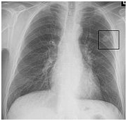
Chest radiograph showing a cancerous tumor in the left lung.
Performing a chest radiograph is the first step if a patient reports symptoms that may be suggestive of lung cancer. This may reveal an obvious mass, widening of the mediastinum (suggestive of spread to lymph nodes there), atelectasis (collapse), consolidation (pneumonia), or pleural effusion. If there are no radiographic findings but the suspicion is high (such as a heavy smoker with blood-stained sputum), bronchoscopy and/or a CT scan may provide the necessary information. Bronchoscopy or CT-guided biopsy is often used to identify the tumor type.
Sputum atypia is associated with an increased risk of lung cancer. Sputum cytologic examination combined with other screening examinations may play an important role in the early detection of lung cancer.
CT scan showing a cancerous tumor in the left lung.
The differential diagnosis for patients who present with abnormalities on chest radiograph includes lung cancer as well as nonmalignant diseases. These include infectious causes such as tuberculosis or pneumonia, or inflammatory conditions such as sarcoidosis. These diseases can result in mediastinal lymphadenopathy or lung nodules, and sometimes mimic lung cancers.


Breast cancer
Breast cancer is a cancer that starts in the breast, usually in the inner lining of the milk ducts or lobules. There are different types of breast cancer, with different stages (spread), aggressiveness, and genetic makeup. With best treatment, 10-year disease-free survival varies from 98% to 10%. Treatment is selected from surgery, drugs (chemotherapy), and radiation.
In the United States, there were 216,000 cases of invasive breast cancer and 40,000 deaths in 2004. Worldwide, breast cancer is the second most common type of cancer after lung cancer (10.4% of all cancer incidence, both sexes counted) and the fifth most common cause of cancer death. In 2004, breast cancer caused 519,000 deaths worldwide (7% of cancer deaths; almost 1% of all deaths).
Breast cancer is about 100 times as frequent among women as among men, but survival rates are equal in both sexes.
Signs and symptoms
The first symptom, or subjective sign, of breast cancer is typically a lump that feels different from the surrounding breast tissue. According to the The Merck Manual, more than 80% of breast cancer cases are discovered when the woman feels a lump. According to the American Cancer Society, the first medical sign, or objective indication of breast cancer as detected by a physician, is discovered by mammogram.Lumps found in lymph nodes located in the armpits can also indicate breast cancer.
Indications of breast cancer other than a lump may include changes in breast size or shape, skin dimpling, nipple inversion, or spontaneous single-nipple discharge. Pain (“mastodynia”) is an unreliable tool in determining the presence or absence of breast cancer, but may be indicative of other breast health issues.
When breast cancer cells invade the dermal lymphatics—small lymph vessels in the skin of the breast—its presentation can resemble skin inflammation and thus is known as inflammatory breast cancer (IBC). Symptoms of inflammatory breast cancer include pain, swelling, warmth and redness throughout the breast, as well as an orange-peel texture to the skin referred to as peau d’orange.
Another reported symptom complex of breast cancer is Paget’s disease of the breast. This syndrome presents as eczematoid skin changes such as redness and mild flaking of the nipple skin. As Paget’s advances, symptoms may include tingling, itching, increased sensitivity, burning, and pain. There may also be discharge from the nipple. Approximately half of women diagnosed with Paget’s also have a lump in the breast.
Occasionally, breast cancer presents as metastatic disease, that is, cancer that has spread beyond the original organ. Metastatic breast cancer will cause symptoms that depend on the location of metastasis. Common sites of metastasis include bone, liver, lung and brain. Unexplained weight loss can occasionally herald an occult breast cancer, as can symptoms of fevers or chills. Bone or joint pains can sometimes be manifestations of metastatic breast cancer, as can jaundice or neurological symptoms. These symptoms are “non-specific”, meaning they can also be manifestations of many other illnesses.
Most symptoms of breast disorder do not turn out to represent underlying breast cancer. Benign breast diseases such as mastitis and fibroadenoma of the breast are more common causes of breast disorder symptoms. The appearance of a new symptom should be taken seriously by both patients and their doctors, because of the possibility of an underlying breast cancer at almost any age.
Causes
The primary risk factors that have been identified are sex,age, childbearing, hormones,a high-fat diet, alcohol intake, obesity, and environmental factors such as tobacco use, radiation and shiftwork.
No etiology is known for 95% of breast cancer cases, while approximately 5% of new breast cancers are attributable to hereditary syndromes In particular, carriers of the breast cancer susceptibility genes, BRCA1 and BRCA2, are at a 30-40% increased risk for breast and ovarian cancer, depending on in which portion of the protein the mutation occurs.
* Personal history of breast cancer: A woman who had breast cancer in one breast has an increased risk of getting cancer in her other breast.
* Family history: A woman’s risk of breast cancer is higher if her mother, sister, or daughter had breast cancer. The risk is higher if her family member got breast cancer before age 40. Having other relatives with breast cancer (in either her mother’s or father’s family) may also increase a woman’s risk.
* Certain breast changes: Some women have cells in the breast that look abnormal under a microscope. Having certain types of abnormal cells (atypical hyperplasia and lobular carcinoma in situ [LCIS]) increases the risk of breast cancer.
* Race: Breast cancer is diagnosed more often in Caucasian women than Latina, Asian, or African American women.
* No physical activity: Women who are physically inactive throughout life may have an increased risk of breast cancer. Being active may help decrease risk.
* Tamoxifen may interact unfavorably with certain antidepressants when used for prevention of breast cancer recurrence.
Diagnosis
This section does not cite any references or sources. Please help improve this article by adding citations to reliable sources. Unsourced material may be challenged and removed. (October 2007)
While screening techniques discussed above are useful in determining the possibility of cancer, a further testing is necessary to confirm whether a lump detected on screening is cancer, as opposed to a benign alternative such as a simple cyst.
In a clinical setting, breast cancer is commonly diagnosed using a “triple test” of clinical breast examination (breast examination by a trained medical practitioner), mammography, and fine needle aspiration cytology. Both mammography and clinical breast exam, also used for screening, can indicate an approximate likelihood that a lump is cancer, and may also identify any other lesions. Fine Needle Aspiration and Cytology (FNAC), which may be done in a GP’s office using local anaesthetic if required, involves attempting to extract a small portion of fluid from the lump. Clear fluid makes the lump highly unlikely to be cancerous, but bloody fluid may be sent off for inspection under a microscope for cancerous cells. Together, these three tools can be used to diagnose breast cancer with a good degree of accuracy.
Other options for biopsy include core biopsy, where a section of the breast lump is removed, and an excisional biopsy, where the entire lump is removed.
Bone tumor
A bone tumor refers to a neoplasic growth of tissue in bone. It can be used for both benign and malignant abnormal growths found in bone, but is most commonly used for primary tumors of bone, such as osteosarcoma. It is may be applied to secondary bone tumors, i.e. metastatic tumors found in bone.
Symptoms
The most common symptom of bone tumors is pain, but many patients will not experience any symptoms, except for a painless mass. Some bone tumors may weaken the structure of the bone, causing pathologic fractures.
